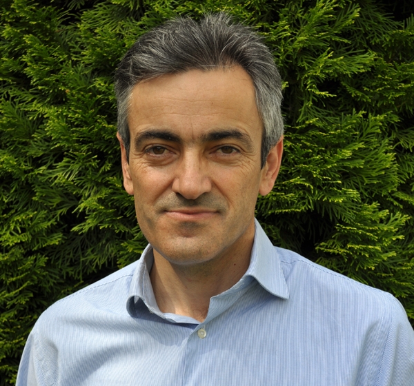
 Savvas Christoforidis
Our group studies the interplay between endocytosis, signaling and exocytosis in endothelial cells and the role they play in vascular physiology.Activated endothelial cells and/or endothelial dysfunction are implicated in the most life-threatening diseases, such as cardiovascular and inflammatory diseases as well as in cancer-angiogenesis. Intriguingly, key vasoactive players of the above pathophysiological processes are stored in specific granules (called Weibel Palade bodies) in endothelial cells. Upon activation of receptors at the cell surface of endothelial cells, Weibel Palade bodies fuse with plasma membrane and release their vasoactive molecules in the blood stream.Our studies have the following aims:1. To shed light on the molecular mechanisms that are responsible for Weibel Palade body trafficking and exocytosis.2. To identify the cell surface receptors and the corresponding signaling cascades that trigger Weibel Palade body exocytosis.3. To track the endocytic routes followed by activated receptors of endothelial cells and the role of these routes in signaling and Weibel Palade body exocytosis.4. To get functional insights into the role of the above processes in vascular physiology and in serious vascular diseases.These studies will shed light on the molecular and functional links between secretion, endocytosis and signalling, as well as on their spatial and temporal coordination. Ultimately, the knowledge generated by these studies will contribute to the design of more effective therapeutic approaches for vascular diseases.
 Vassilis Douris
The role of specific genes and mutations in insecticide resistance We employ CRISPR/Cas9 genome modification in Drosophila to validate candidate mutations through a reverse genetics approach (reviewed in Douris et al., 2020 Pestic Biochem Physiol). We also perform functional expression of candidate resistance genes (P450s) to investigate their ability to metabolize insecticides in vivo (Tsakireli et al., 2019 Pestic Biochem Physiol; Riga et al., 2020 Insects). We have previously investigated resistance against several chemistries, and currently focus on mechanisms of target-site and metabolic resistance to avermectins. Synergistic interactions among different resistance mechanisms While epistatic effects among genes involved in co-existing resistance mechanisms are postulated from field data, we were able to show a clear synergistic interaction between target-site mutations and metabolic resistance alleles in an engineered Drosophila system devoid of confounding genetic effects, regarding resistance to pyrethroids (Samantsidis, Panteleri et al., 2020 Proc Roy Soc B). Furthermore, we were able to assess potential fitness disadvantage conferred by the coexistence of multiple alleles. We now investigate a similar framework with different molecular targets and insecticide chemistries, involving resistance to avermectins. Novel resistance mechanisms and new potential compounds Having contributed to the establishment of a novel class of proteins (chemosensory proteins, CSPs) as potential players in pyrethroid resistance in mosquitoes (Ingham et al., 2020 Nature) we seek to investigate the role of CSPs as insecticide binding proteins with potential contribution in metabolic resistance or sequestration in vivo. Currently, we are functionally expressing candidate lepidopteran CSPs in insect cell (baculovirus) systems and assess their potential within the framework of iNEXT / INSTRUCT-ERIC in collaboration with the NKI protein facility (The Netherlands). We seek to identify optimal binding modules that could inform rational design of new potential compounds and/or synergists for insecticide formulations. Investigation of resistance mechanisms and their interactions through genome editing and genetic engineering in Drosophila (Douris et al., 2020 Pestic Biochem Physiol). Research aims: Our lab studies the molecular mechanisms underlying insecticide resistance. Our goal is:1. To understand how specific mutations and gene regulation shape resistance phenotypes in natural populations2. To investigate synergistic interactions among different mechanisms and their impact in fitness3. To explore novel resistance mechanisms and develop applications towards potential new compounds and synergists We work with Drosophila models and insect cell culture and use molecular biology, genomics, transcriptomics, and proteomics approaches, CRISPR/Cas9 genome modification and functional protein expression. We expect to create a collaborative network of scientific interactions, promoting scientific advancement and social outreach.
 Michaela Filiou
Our lab studies the molecular mechanisms of neuropsychiatric disorders. Our goal is:• To understand how mitochondria, organelles that fuel cells with energy, shape stress- and anxiety- related behaviors• To identify molecular signatures and candidate biomarkers for neuropsychiatric phenotypes and treatment responses• To explore the potential of mitochondria as therapeutic targets for neuropsychiatric disordersWe work with mouse models, patient samples and cell culture and we use behavioral biology, proteomics/metabolomics approaches, protein biochemistry and pharmacology. Our vision is to create a collaborative environment, fostering scientific growth and communication.
 Maria Georgiadou
Lung cancer is the leading cause of morbidity and mortality worldwide. Unfortunately, most lung cancers present as stage IV attributed to metastatic disease, which is associated with increased symptom burden and poor survival. Novel and potent therapy options have transformed the treatment of some subsets of lung cancer; nevertheless, the clinical benefit is still limited to a minority of patients reflecting the need to better understand the underlying biology of the disease. This limited response is mainly attributed to therapeutic resistance, which is manifested as local or distant disease recurrence. Stiffness is a biophysical property of the ECM that modulates cellular functions, including proliferation, invasion, differentiation, and it may affect therapeutic responses. The ability of the cells to sense tissue stiffness and convert these mechanical cues into intracellular changes is termed mechanotrasduction. While it is well established that ECM stiffness influences the behaviour of normal cells the role of matrix stiffness in transformed cells – and more precisely in lung cancer cells – remains largely unclear. We are using hydrogels with tuneable stiffness to study the impact of ECM rigidity on lung cancer cells growth, motility, response to drug treatment and effects on cell metabolism. Figure 1. (A) Schematic of the fabrication of hydrogels with tuneable stiffness; (B) Representative images of HCC827 lung cancer cells plated on soft (left) or stiff (right) hydrogels and stained for the actin marker phalloidin and active ?1-integrin antibody (12G10). Research aims: To understand how extracellular matrix stiffness regulates lung cancer cell growth, migratory behaviour and drug resistance To identify novel therapeutic vulnerabilities in lung cancer cells treated with targeted therapies
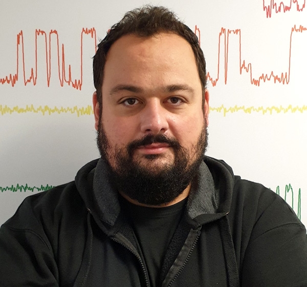 Christos G. Gkogkas
The complex polygenic nature of brain disorders, such as neurodevelopmental disorders (Autism Spectrum Disorders, fragile X syndrome, intellectual disability) neuropsychiatric disorders (depression, schizophrenia) and neurodegenerative disorders (Parkinson's disease, Alzheimer's disease) along with the plethora of contributing non-genetic factors have impeded our efforts to understand and treat them. By studying converging signalling, molecular and cellular pathways, we can shed fresh light on the causality of brain disorders.Proteins catalyse most of the reactions in the cell on which life depends. Translational control is defined as the sum of regulatory events that dictate changes in protein synthesis, per mRNA, per unit of time, and it is a powerful means to alter protein abundance. Our lab is particularly interested in understanding the molecular and signalling mechanisms of translational control in the brain and how they control complex brain functions and behaviours, such as learning, memory, social interactions, anxiety and fear and how they affect brain health (e.g.neurodegeneration). Our studies have the following aims: Investigating Post-Transcriptional Translational Control: The lab aims to elucidate the mechanisms of post-transcriptional regulation of gene expression in the brain, focusing on how signalling and RNA-binding proteins modulate mRNA translation. Utilizing Human Pluripotent Stem Cells: The lab seeks to leverage human pluripotent stem cell-derived brain organoids to study the development and function of human neural circuits. This approach will provide insights into the cellular and molecular pathways that are disrupted in neurological diseases. Developing Rodent Models of Disease: We aim to develop and characterize rodent models that accurately mimic human neurobiological disorders, such as autism. These models will facilitate the investigation of disease mechanisms and the testing of potential therapeutic compounds. Innovating Therapeutic Strategies: We aim to explore novel therapeutic strategies targeting the dysregulation of protein synthesis in neurological disorders. By developing genetic and pharmacological tools, we aim to restore normal protein synthesis and ameliorate disease symptoms.
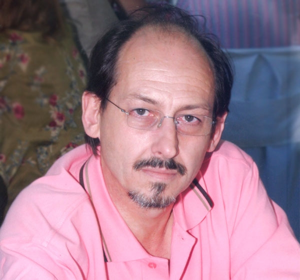 Evangelos Kolettas
NF-κB signalling pathways and IKK/NF-κB-miRNA regulatory network in DNA damage and inflammation impacting on senescence and cancerNF-κB transcription factors (TFs) are critical regulators of pro-inflammatory/stress-like responses, and their immediate upstream signaling components are aberrantly expressed and/or activated in pulmonary diseases and in non-small cell lung cancer (NSCLC), with an unfavourable prognosis for patient survival. Activation of NF-κB is achieved by two main signalling pathways: An IKKβ-mediated canonical NF-κB signalling pathway involving the nuclear translocation of c-Rel/p50 or p65/50 heterodimes where they regulate target gene expression, and an IKKβ-mediated non-canonical or alternative NF-κB signalling pathway involving the nuclear translocation of p52/RelB heterodimers to regulate a distinct set of NF-κB target gene expression.Using in vitro cell systems and in vivo novel mouse lung cancer models, we study the:1. Mechanisms of action of canonical IKKβ/NF-κB in NSCLC2. Role of the regulatory NF-κB-miRNA network in NSCLC3. Mechanism of action of IKKα in NSCLC4. Mechanisms of action of IKKα in oncogene induced senescenceIdentifying novel regulators of DNA damage, inflammation and cancer by employing domain-specific CRISPR/Cas9 screensWe will employ a functional kinase- or a transcription factor-specific CRISPR/Cas9 lentiviral library to ablate protein kinases or TFs and identify novel regulators involved in DNA damage and inflammation impacting on cancer.
 Petros Marangos
The process of Meiosis in mammalian female germ cells is initiated in the ovaries before birth. The total number of oocytes is established in the ovaries during embryogenesis when the germ cells enter Meiosis and remain arrested at Prophase. The quiescent state of Prophase arrest may last for years, and in humans, Prophase arrest may last for up to 50 years. From puberty, oocytes receive a stimulus to grow and obtain the competency to resume Meiosis. This protracted state of arrest renders oocytes extremely susceptible to accumulating environmental or endogenous insults, which may affect the genetic integrity of the female gametes and therefore the genetic integrity of the outcoming embryo. Furthermore, a long-lasting state of arrest affects oocyte physiology and dynamics which lead to the well documented age-related infertility. Fertility and the physiological embryonic development depend largely on the correct regulation of the cell cycle during gametogenesis and early embryonic development. Our Lab focuses on the examination of cell cycle regulation and the DNA damage response during mammalian oocyte and preimplantation development. The specific research activities of the Developmental & Reproductive Biology Lab are: Cell cycle regulation in mammalian oocytes and pre-implantation embryos. DNA damage response in mammalian oocytes and pre-implantation embryos. Role and function of oocyte-specific regulators that show oncogenic expression. Mechanisms of oocyte aging and the relation of oocyte aging with chromosomal abnormalities and DNA damage. Effects of nutritional and anxiety disorders on the development of mammalian oocytes.
 Carol Murphy
Vasculogenesis, is a fundamental developmental process, during which mesodermal progenitors are differentiated to endothelial cells (ECs). The initial vessel plexus, formed from ECs only, further matures via the process of vascular myogenesis, during which mural cells (MCs), such as pericytes (PCs) and vascular smooth muscle cells (vSMCs), are recruited to the vessels via cross-talk with ECs differentiating locally. Such mature vessels consisting from ECs and PCs/SMCs can form new vessels using spouting in a process called angiogenesis. Many molecules participate in regulating vessel morphogenesis during early development (vasculogenesis) or at later stages of development and postnatally (angiogenesis). Interestingly, VEGFA appears to regulate both processes though in vasculogenesis it achieves the differentiation of mesodermal to vascular progenitor cells (VPCs) and further to ECs whereas in angiogenesis it generates new vessels by sprouting of ECs. Thus, VEGF signalling generates the excessive angiogenesis seen in angiogenic diseases and particularly in tumours. Likewise, VEGF cascades are required for the vasculogenic vessel engineering in Regenerative Medicine. Therefore, a detailed knowledge of VEGF signalling that regulates both angiogenesis and vasculogenesis is needed. However, at present there is almost no information about the vasculogenic VEGF cascades. Vessel engineering is critical for the success of RM because the speed by which the inoculated cells are vascularised by the host is important. Delayed vascularisation of the implant results in low cell survival rates. Thus, ideally the tissue engineered construct (TEC) should be already vascularised in vitro prior to implantation in vivo. We have already initiated a programme for vessel engineering based on reprogramming human fibroblasts to generate human induced pluripotent stem cells (hiPSCs) and subsequent differentiation of hiPSCs, via mesodermal intermediates, to VPCs and then to ECs in chemically defined media (CDM) or to PCs/vSMCs. Whereas this is a step forward, there are many gaps in our knowledge that need to be addressed. Indeed, the mesodermal VEGFR2+ intermediate cell populations and VPCs are not well characterised and almost nothing is known about the VEGF-A-induced signalling cascades that differentiate/commit mesodermal intermediates to VPCs and then to ECs. Using a combination of cutting-edge technologies, we will explore the signalling pathways of VEGF-A responsible for vasculogenesis, the de novo emergence of endothelial cells from mesoderm (see aims). Recently, we have extended our research into the role of endothelial cells in neurodegenerative diseases, using Parkinson’s Disease as a model system. We are establishing a PD NeuroVascular Unit combining all relevant cells (astrocytes, microglia, dopaminergic neurons, endothelial cells and pericytes) in a microfluidic platform to investigate the cell-cell communication and alterations in this system. Additionally, this system will be used for preclinical studies on treatments for PD. Aims Determining the VEGF-induced circuit regulating vasculogenesis during vessel engineering by employing phosphoproteomics, single cell RNAseq, single cell ATACseq and bioinformatics. Evaluating the involvement of flow/shear stress and Mural cells in the differentiation of hiPSCs to ECs via mesodermal intermediates in microfluidic platforms. Establishing a preclinical model of PD using human induced pluripotent stem cells (hiPSCs) from PD patients and isogenic controls to investigate the NeuroVascular Unit in PD.
 Frank Fackelmayer
Our group is interested in epigenetics and the functional architecture of the eukaryotic cell nucleus, and their role in human development, health and disease. It is generally accepted that chromatin is organized in topologically associated loop domains that are formed and maintained to provide the intricate infrastructure needed to properly regulate gene expression and DNA replication. We study how this high-level organization of chromatin is regulated by architectural proteins of the cell nucleus and their epigenetic modifications. One focus of our research is the Scaffold Attachment Factor A (SAF-A), an essential component of the nuclear scaffold/matrix that is involved in folding chromatin to functionally independent units of gene expression. Importantly, SAF-A also associates with PRMT1, a member of the epigenetic family of protein arginine methyltransferases, and apparently recruits the enzyme to strategically important regions of the genome to contribute to the epigenetic regulation of the local gene accessibility and activity. Image: SAF-A is essential for the functional architecture of the cell nucleus. Left panels show that full length wildtype SAF-A and bulk chromatin co-localize in the nucleus, while the right panels show how a dominant negative construct of SAF-A, called C280, leads to a global collapse of chromatin structure. Each panel shows one transfected cell and no-transfected neighboring cells for control. Note how C280 and collapsed chromatin are mutually exclusive. Our ongoing research has the following aims: * understand how SAF-A affects gene expression through regulation of chromatin folding * establish live-cell methods to study the connection of nuclear structure and cell physiology * delineate cellular pathways directly and indirectly affected by SAF-A * decipher the functional consequences of this regulation on cell physiology and differentiation * elucidate how arginine methylation by PRMTs is involved in regulating chromatin organization Taken together, the ongoing studies will shed light on fundamental aspects of nuclear architecture, and will lead the way to harnessing this new knowledge for the rational design of novel applications in medicine.
 Dimitris Beis
Associate Professor of Biological Chemistry, Faculty of Medicine, University of Ioannina. Abstract of your research field My research interests are in the areas of Biochemistry, Cellular and Developmental Biology. My lab is using zebrafish to model Human Disease and identify novel bioactive compounds. The main focus of my lab is on cardiovascular disease and angiogenesis. We have been able to identify the significance of intracardiac flow dynamics during the development of the heart for proper cardiac valve development. We recently expanded our strategy to study the mechanisms of cardiovascular regeneration exploiting the extraordinary regenerative potential of zebrafish. We showed that zebrafish can regenerate their cardiac valves and identified the Notch and TGF? signaling pathways as key regulators for the first steps of this process. We now study the role of the innate immune response during cardiac regeneration. Photo description A) Zebrafish are a valuable model for Biomedical Research. B) Confocal 3D reconstruction of an adult zebrafish heart, outlining the heart vessels (coronary network) using the fli1:GFP transgenic line. C) Confocal 3D reconstruction of an adult zebrafish heart with the atrioventricular and aortic valves stained with an elastin antibody. Research aims: Identify novel genes causing cardiovascular disease, exploiting GWAS and variants of unknown significance from Human Genetic studies Identify the role of the innate immune response during cardiac regeneration Identify novel bioactive compounds from natural products and other complex extracts
 Dimitrios I. Fotiadis
The Unit of Medical Technology and Intelligent Information Systems is a highly innovative and self-contained research unit that resides at the Dept. of Material Science & Engineering of the University of Ioannina. The unit is strongly activated in the fields of Biomedical Engineering and development of Intelligent Information systems. It has an internationally acknowledged excellence in conducting high quality scientific research and developing innovative Information Technology (IT) applications, products and services. The unit is co-ordinated by Prof. Dimitrios I. Fotiadis, who is Professor of “Biomedical Engineering” at the Dept.of Materials Science and Engineering of the University of Ioannina and staffed with qualified scientists, researchers and other IT personnel. Figure Legend: Research activities of the Unit Main Research Multi-scale Modelling of Biological Tissues and Systems The Unit of Medical Technology and Intelligent Information Systems has shown a unique expertise in multiscale modelling of biological tissues and systems for over the last two decades. The scope of research is wide, dealing with numerical modelling of: a) Numerical modelling of cardiovascular disease, b) Computational modelling of bone pathologies, c) glycaemic control in diabetic patients, and iv) Modelling of hearing function in middle and inner ear. Biomedical and Genetic Data processing The Unit of Medical Technology and Intelligent Information Systems is active in several fields of bioinformatics, such as: (i) Sequence classification based on pattern mining, (ii) Cis/trans isomerization of the peptide bond using data mining, (iii) Protein fold recognition using hidden Markov model and (iv) Evolution of oral cancer based on the integration of heterogeneous data and data analysis methods. Intelligent information systems This broad domain includes the development and validation of state-of-the-art telemedicine systems for the monitoring of patients with chronic diseases, patients rehabilitating after skeletal traumas or sports injuries, and people needing special care such as pregnant women or subjects with phobias. Wearable Systems The research objective is twofold: a) computational modeling of ultrasonic propagation in healing and osteoporotic long bones to monitor the evolution of osteoporosis and fracture healing, b) the development of a biomechanical methodology for the description of the bone-implant relation.
 Costas Papaloukas
Bioinformatics combines Biology and Computer Science and can be divided into three main subfields: a) the development of new algorithms and methods for the assessment of large data sets, b) the analysis and interpretation of various types of data, including biological sequences, protein structures and domains, as well as clinical and biomedical ones, and c) the development and implementation of tools that allow efficient access and management of different types of information. In the Bioinformatics laboratory, special emphasis is given on independent research in up-to-date areas, such as the computational analysis of proteins and biomolecules in general, the development of computational techniques for better studying biological systems, including environmental and aquatic ones, as well as biomedical technology. Clustering of gene expression data using SOMs Research aims: Biomedical data storage & management Processing & analysis of DNA & protein sequences High-throughput data analysis Diagnosis of pathological tissues (based on gene expression profiles) Medical decision support systems
EMERITUS
 Spyros Georgatos
BiographySpyros D. Georgatos is a member of BRI-FORTH since 2002. He holds a Medical Degree from the University of Athens, Greece (1980) and a Ph.D. in Biology from Yale University, USA (1985). He post-docked at Caltech and Rockefeller University in the US and served as a group leader in the Cell Biology Program of the European Molecular Biology Laboratory (EMBL) in the period 1990-1996. In 1999 he was elected an EMBO member, while serving as an Associate Professor at the University of Crete, School of Medicine, Greece. He is Head of the Laboratory of Biology at the Ioannina University, School of Medicine. Prof. Georgatos has published one book and numerous research articles (over 3.000 citations). His recent interests include stem cell differentiation and division, as it relates to nuclear structure and function. His administrative work includes the establishment of one of the first Graduate Programs in Cell and Molecular Biology in Greece (1998), service at the Scientific Board of the Hellenic Pasteur Institute (2001-2004) and management of several research grants.
 Theodore Fotsis
Vasculogenesis, is a fundamental developmental process, during which mesodermal progenitors are differentiated to endothelial cells (ECs). The initial vessel plexus, formed from ECs only, further matures via the process of vascular myogenesis, during which mural cells (MCs), such as pericytes (PCs) and vascular smooth muscle cells (vSMCs), are recruited to the vessels via cross-talk with ECs differentiating locally. Such mature vessels consisting from ECs and PCs/SMCs can form new vessels using spouting in a process called angiogenesis. Many molecules participate in regulating vessel morphogenesis during early development (vasculogenesis) or at later stages of development and postnatally (angiogenesis). Interestingly, VEGFA appears to regulate both processes though in vasculogenesis it achieves the differentiation of mesodermal to vascular progenitor cells (VPCs) and further to ECs whereas in angiogenesis it generates new vessels by sprouting of ECs. Thus, VEGF signalling generates the excessive angiogenesis seen in angiogenic diseases and particularly in tumours. Likewise, VEGF cascades are required for the vasculogenic vessel engineering in Regenerative Medicine. Therefore, a detailed knowledge of VEGF signalling that regulates both angiogenesis and vasculogenesis is needed. However, at present there is almost no information about the vasculogenic VEGF cascades. Vessel engineering is critical for the success of RM because the speed by which the inoculated cells are vascularised by the host is important. Delayed vascularisation of the implant results in low cell survival rates. Thus, ideally the tissue engineered construct (TEC) should be already vascularised in vitro prior to implantation in vivo. We have already initiated a programme for vessel engineering based on reprogramming human fibroblasts to generate human induced pluripotent stem cells (hiPSCs) and subsequent differentiation of hiPSCs, via mesodermal intermediates, to VPCs and then to ECs in chemically defined media (CDM) or to PCs/vSMCs. Whereas this is a step forward, there are many gaps in our knowledge that need to be addressed. Indeed, the mesodermal VEGFR2+ intermediate cell populations and VPCs are not well characterised and almost nothing is known about the VEGF-A-induced signalling cascades that differentiate/commit mesodermal intermediates to VPCs and then to ECs. Using a combination of cutting-edge technologies, we will explore the signalling pathways of VEGF-A responsible for vasculogenesis, the de novo emergence of endothelial cells from mesoderm (see aims). Recently, we have extended our research into the role of endothelial cells in neurodegenerative diseases, using Parkinson’s Disease as a model system. We are establishing a PD NeuroVascular Unit combining all relevant cells (astrocytes, microglia, dopaminergic neurons, endothelial cells and pericytes) in a microfluidic platform to investigate the cell-cell communication and alterations in this system. Additionally, this system will be used for preclinical studies on treatments for PD. Aims Determining the VEGF-induced circuit regulating vasculogenesis during vessel engineering by employing phosphoproteomics, single cell RNAseq, single cell ATACseq and bioinformatics. Evaluating the involvement of flow/shear stress and Mural cells in the differentiation of hiPSCs to ECs via mesodermal intermediates in microfluidic platforms. Establishing a preclinical model of PD using human induced pluripotent stem cells (hiPSCs) from PD patients and isogenic controls to investigate the NeuroVascular Unit in PD.
 Anastasia S. Politou
 is a member of BRI-FORTH since 2007. She holds a B.Sc. in Chemistry from the University of Athens, Greece and M.Sc. and PhD degrees in Chemistry from New York University, USA. She worked as a post-doctoral fellow in the Structural Biology and Biocomputing Program of the European Molecular Biology Laboratory (EMBL), as a Visiting Associate Professor in the Chemistry Dept., University of Crete and as a Research Fellow in the National Institute for Medical Research, Medical Research Council, Mill Hill, London, UK. Since 2001 she has been working as an Assistant Professor at the Ioannina University, School of Medicine. A.S. Politou has published 35 research articles (over 1.080 citations). Her recent research interests include the study of the structure and dynamics of chromatin-related proteins and protein complexes. Her administrative work includes the establishment and management of the first High Resolution NMR facility in Greece (University of Crete, 1996-1999) and of the DNA Microarray facility at the University of Ioannina and the management of several research grants.
Anastasia S. Politou
Anastasia S. Politou is a member of BRI-FORTH since 2007. She holds a B.Sc. in Chemistry from the University of Athens, Greece and M.Sc. and PhD degrees in Chemistry from New York University, USA. She worked as a post-doctoral fellow in the Structural Biology and Biocomputing Program of the European Molecular Biology Laboratory (EMBL), as a Visiting Associate Professor in the Chemistry Dept., University of Crete and as a Research Fellow in the National Institute for Medical Research, Medical Research Council, Mill Hill, London, UK. Since 2001 she has been working as an Assistant Professor at the Ioannina University, School of Medicine. A.S. Politou has published 35 research articles (over 1.080 citations). Her recent research interests include the study of the structure and dynamics of chromatin-related proteins and protein complexes. Her administrative work includes the establishment and management of the first High Resolution NMR facility in Greece (University of Crete, 1996-1999) and of the DNA Microarray facility at the University of Ioannina and the management of several research grants.


